


Reconstructions Involving Multiple Modalities
-
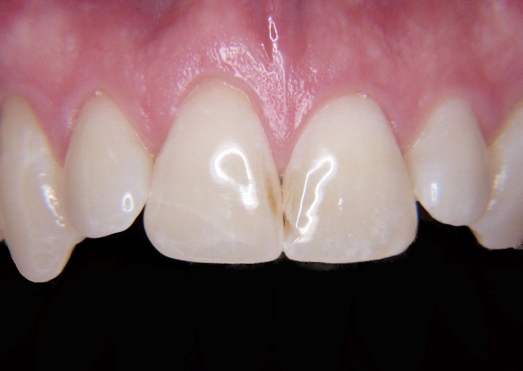
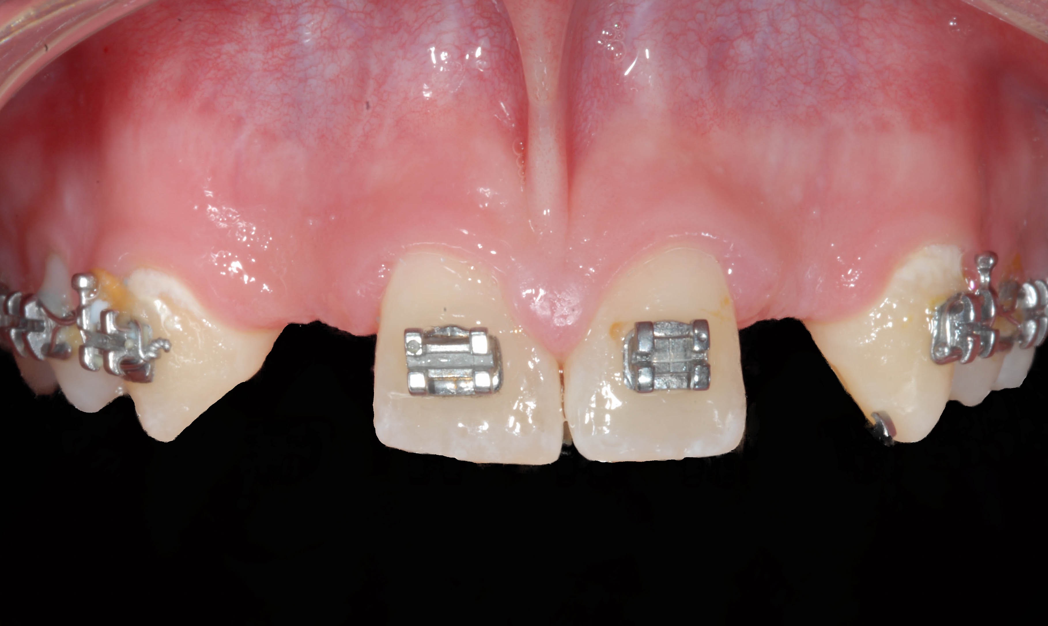
Anterior tooth requiring removal due to resorption of the root from previous trauma.
-
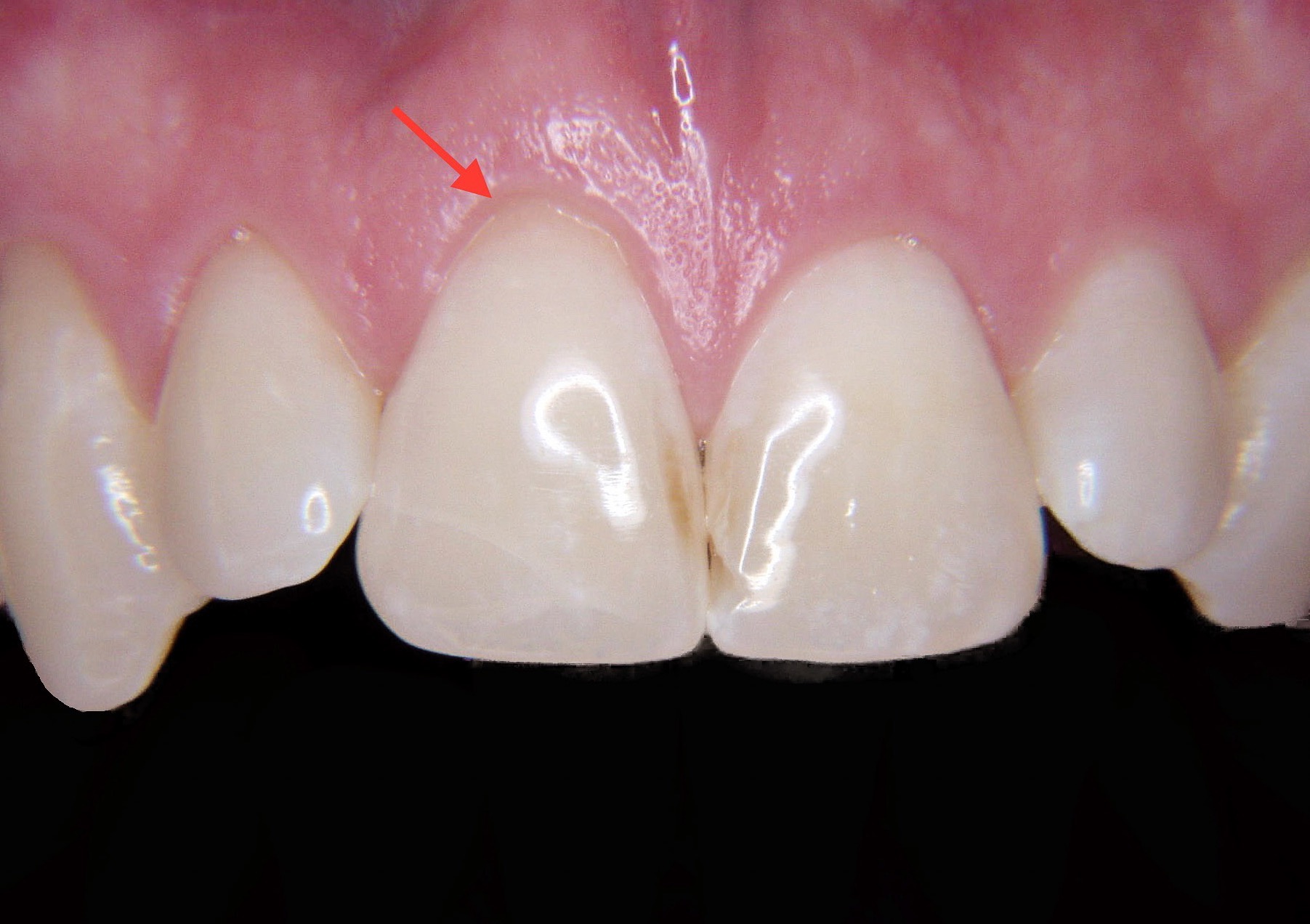
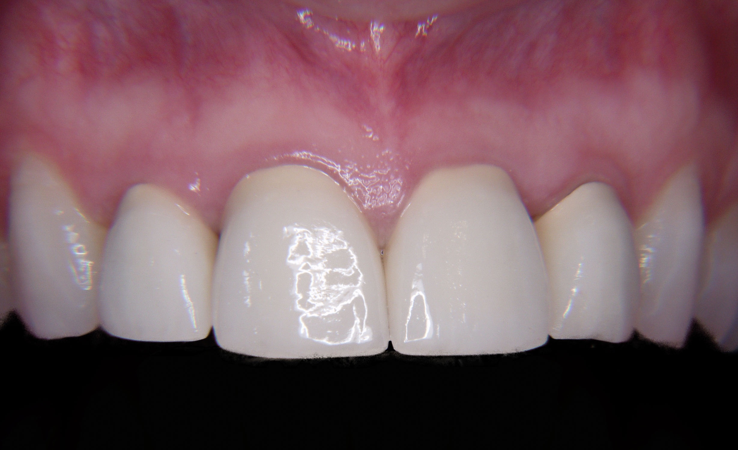
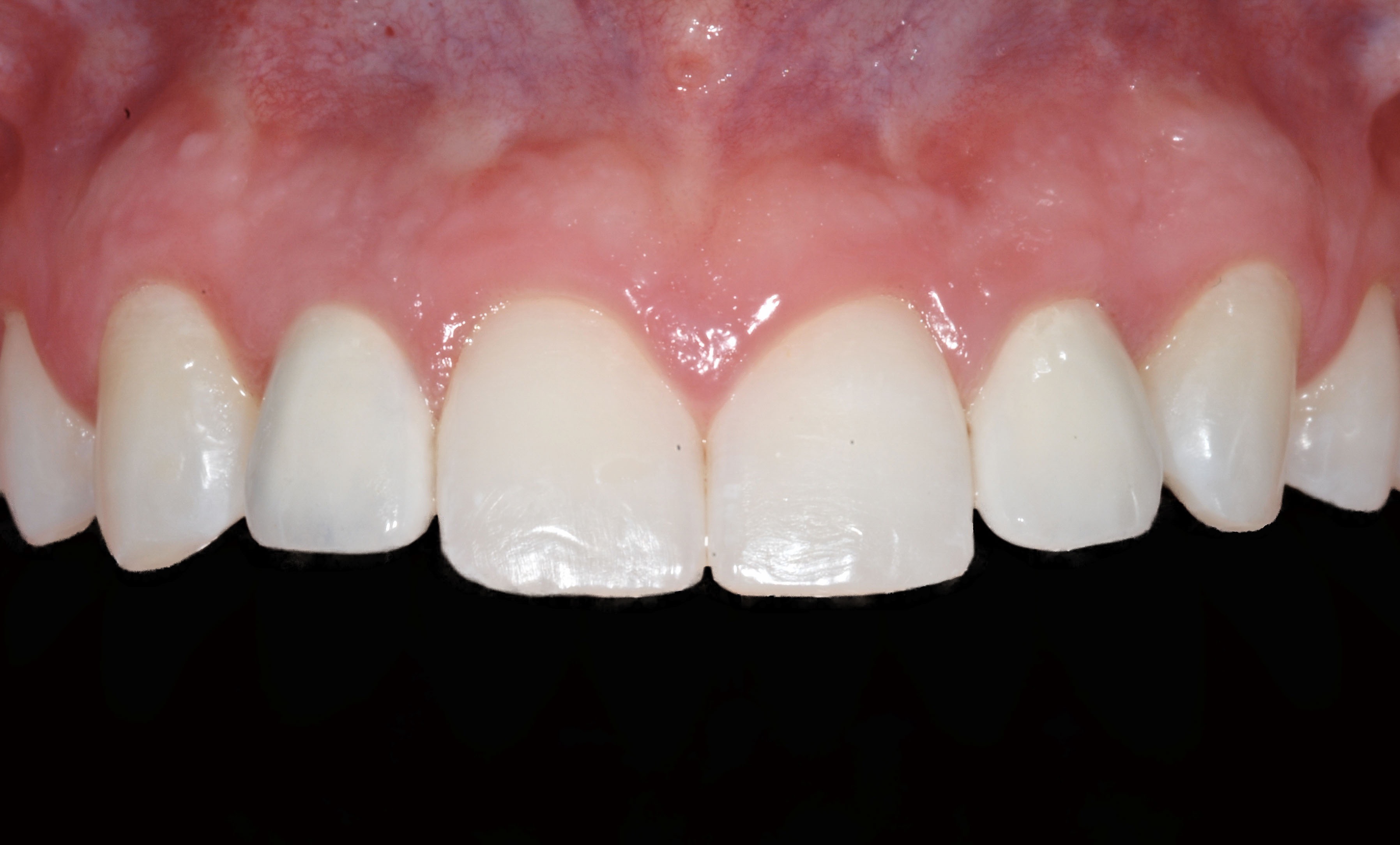
Patient did not like the recession as the tooth looked longer the the adjacent central incisor (arrow).
-
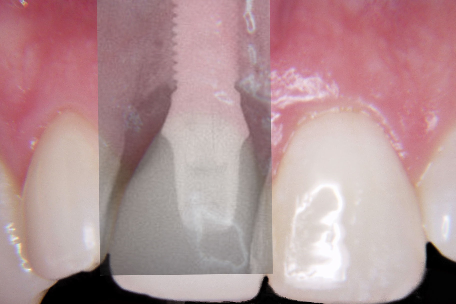
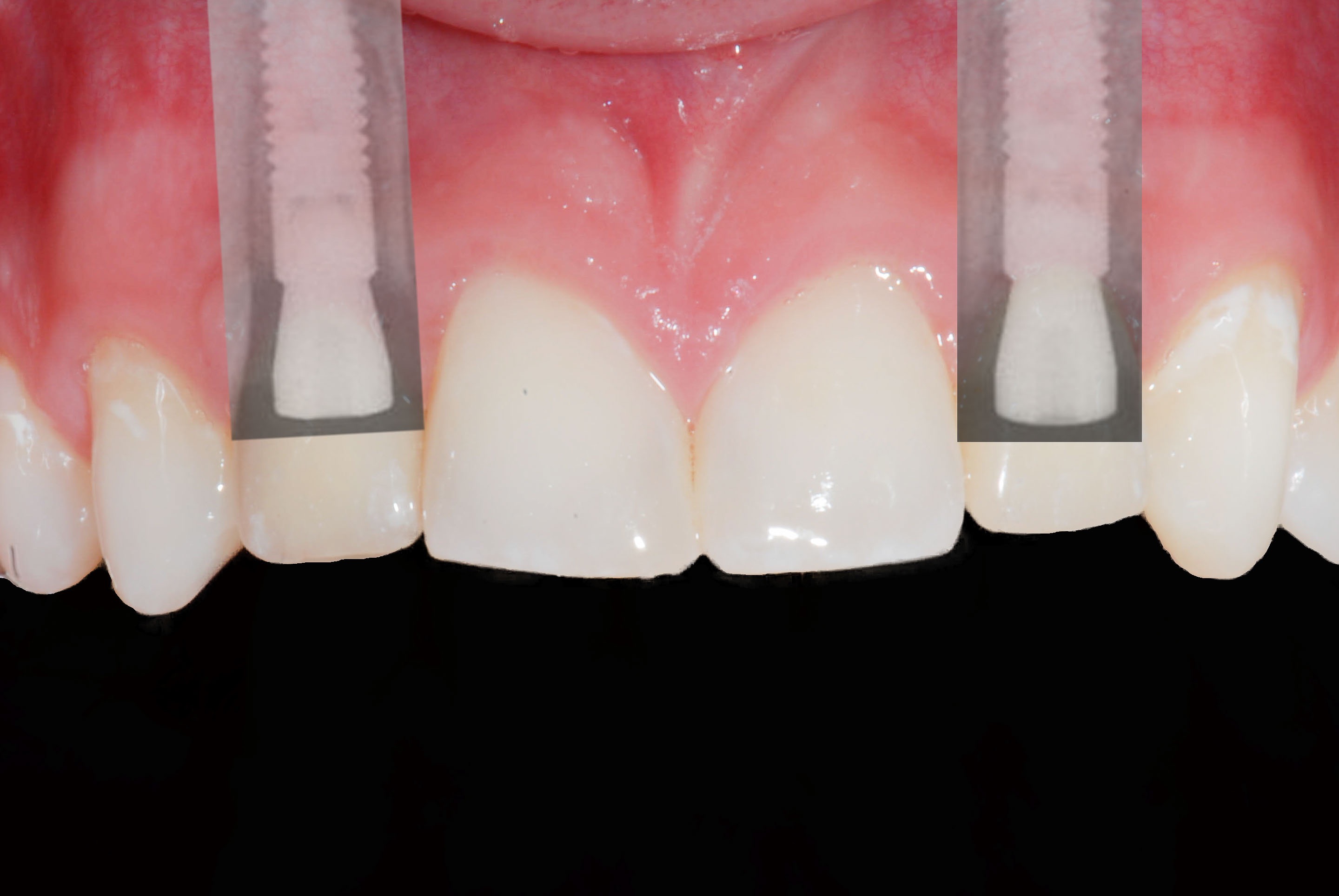
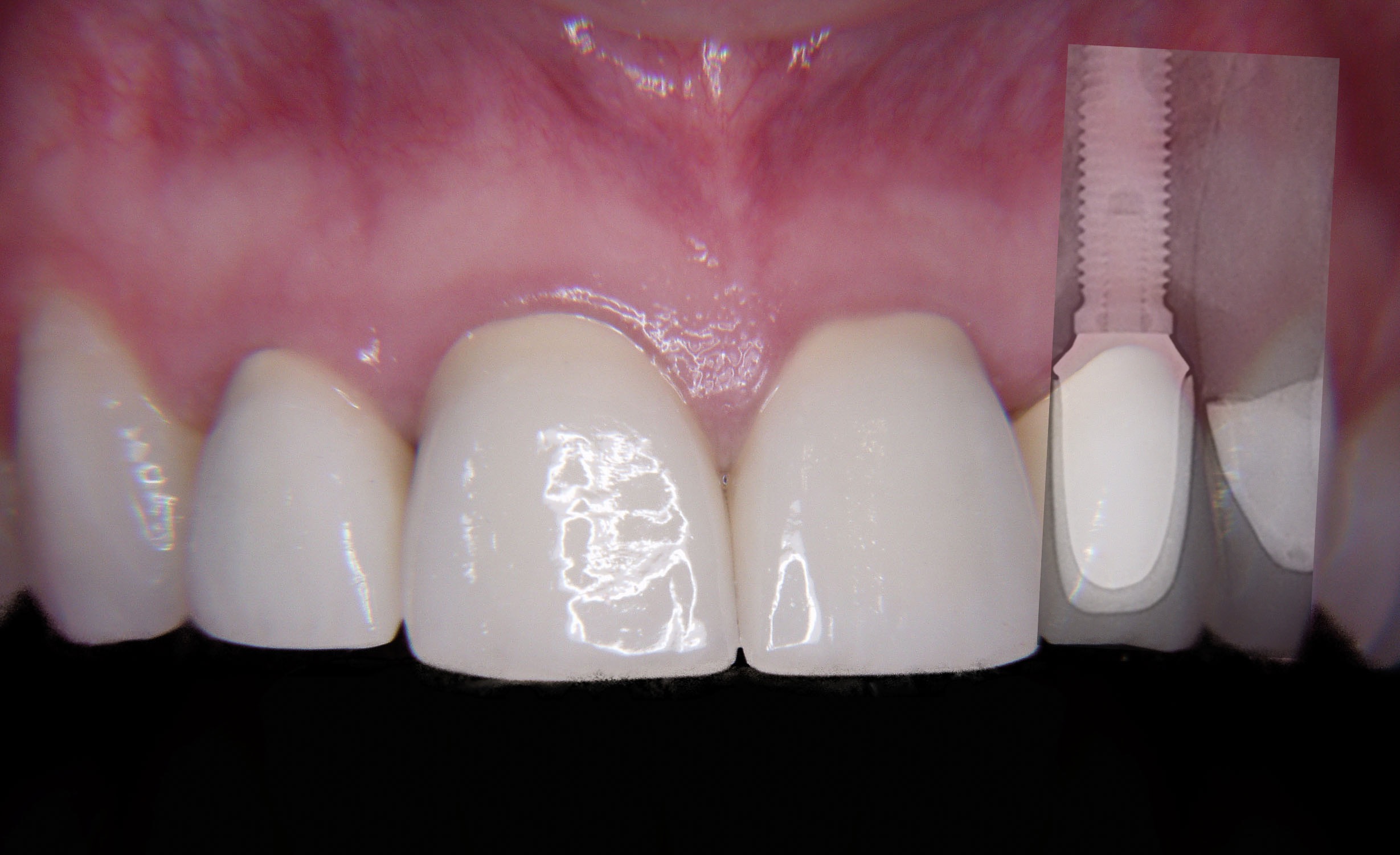
Radiographic overlay showing implant which was immediately placed at the time the tooth was removed.
-
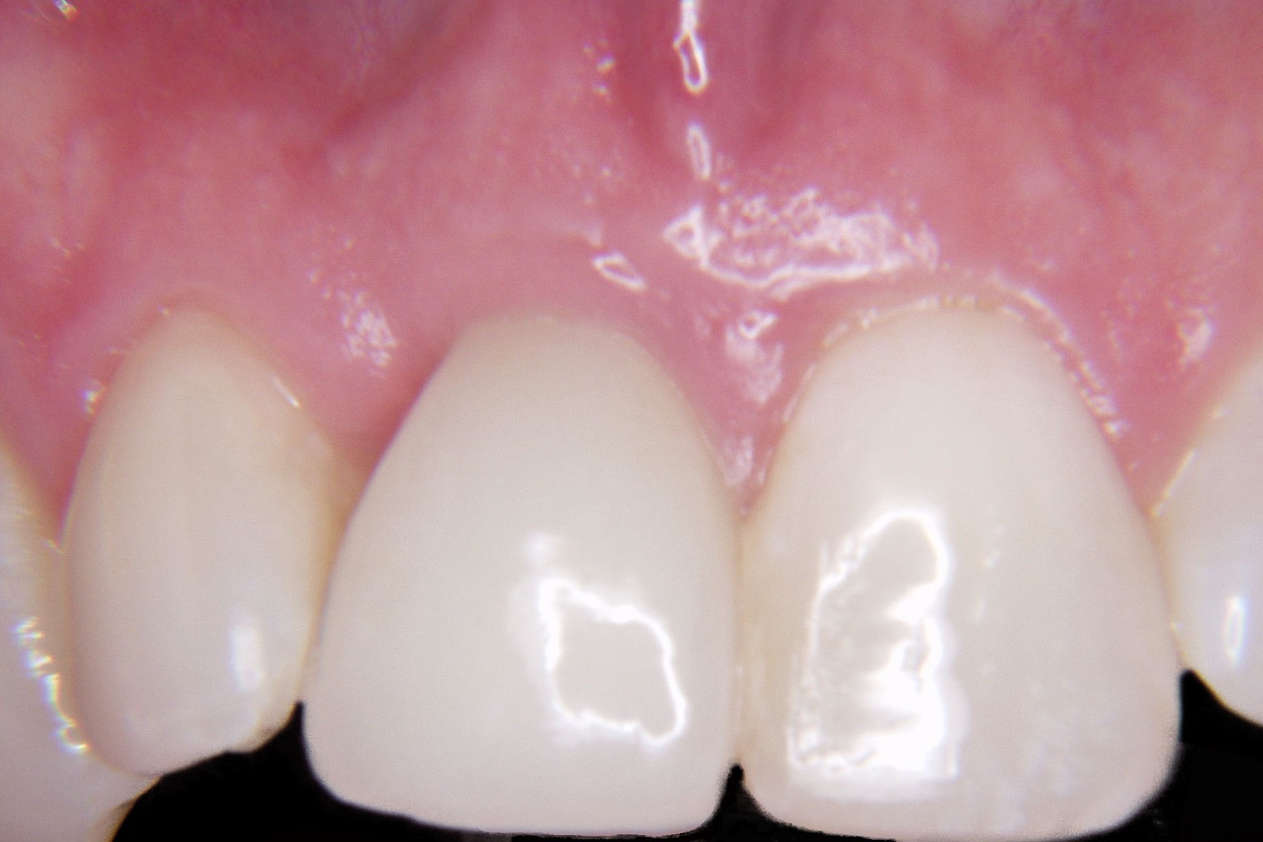
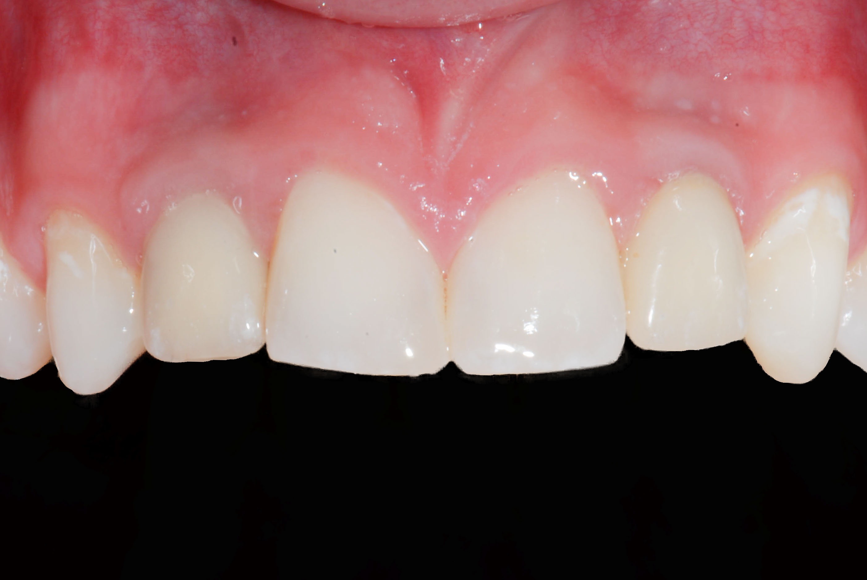
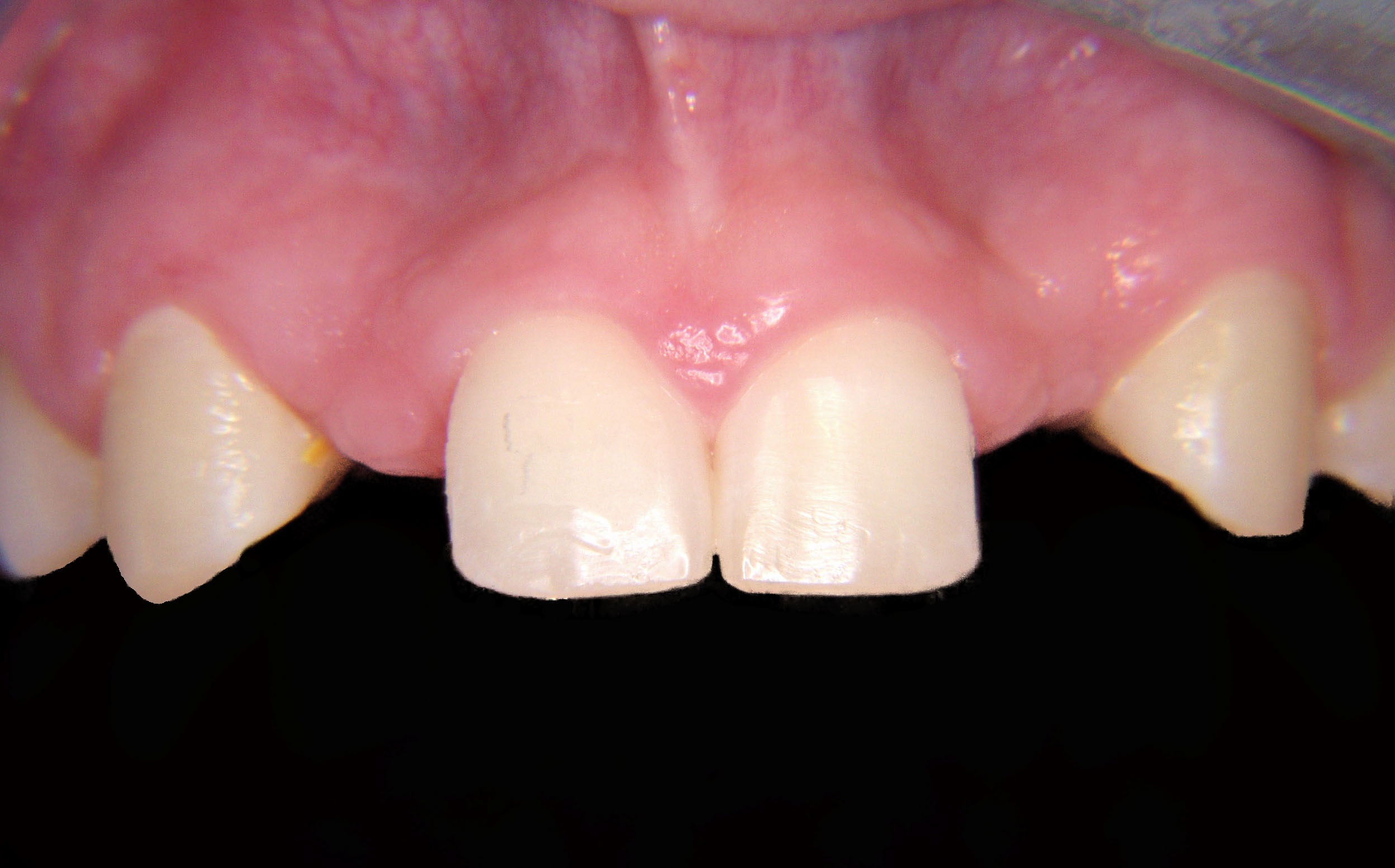
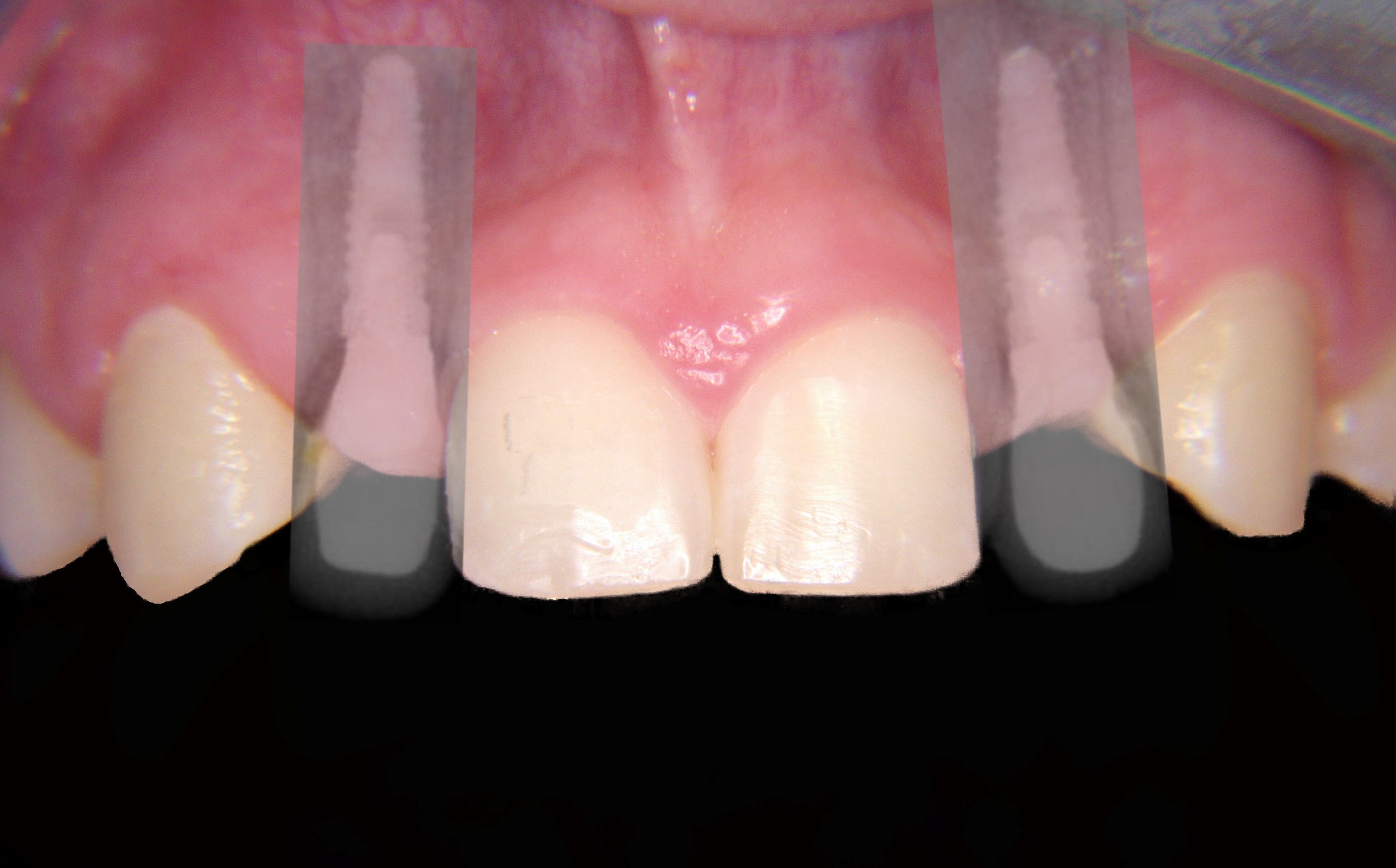
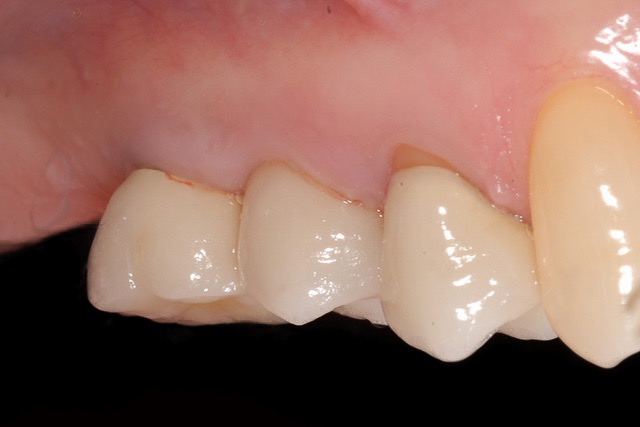
Final restoration after 3 months for the implant to "integrate" with the bone.
-
-
-
-
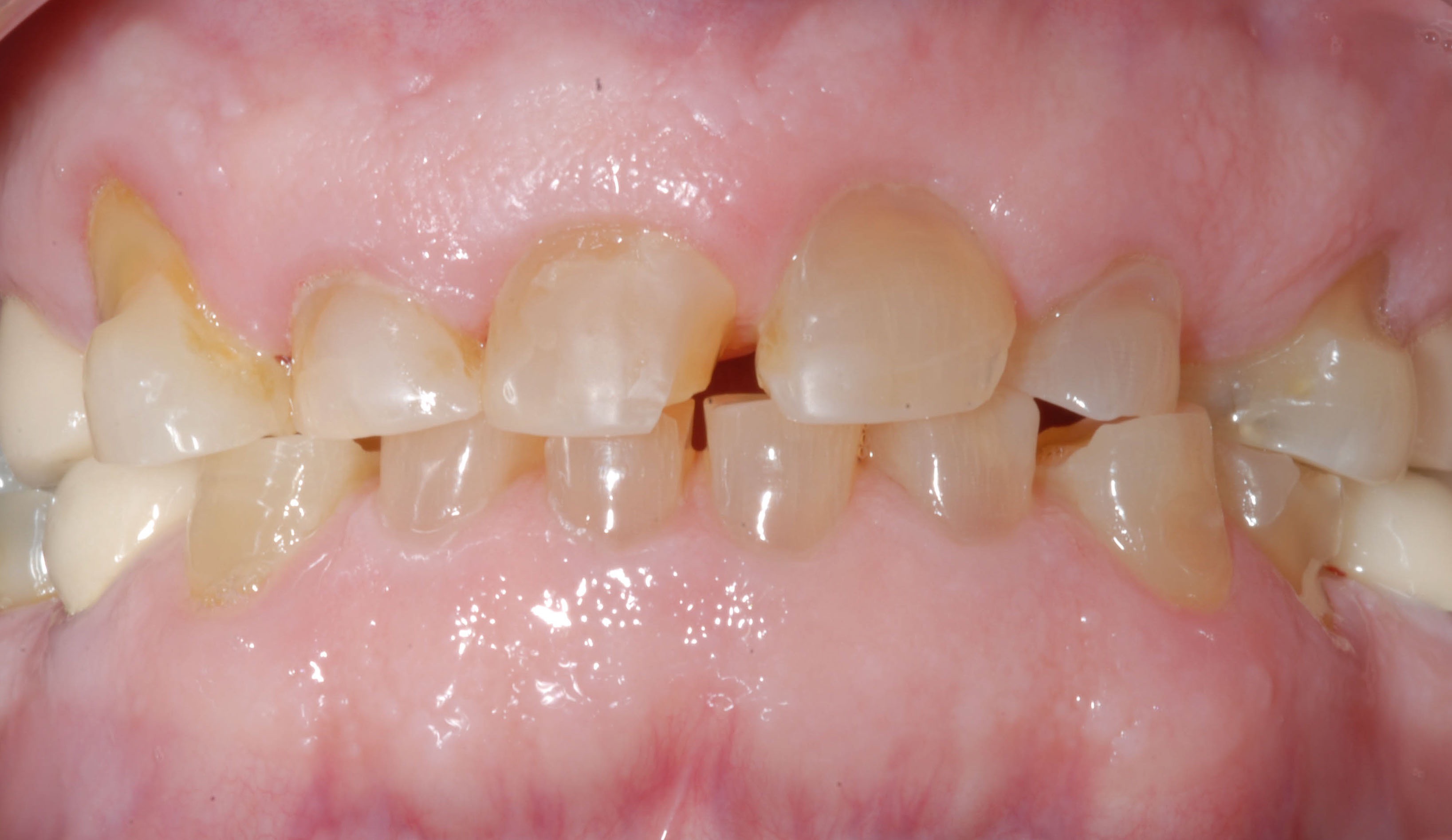
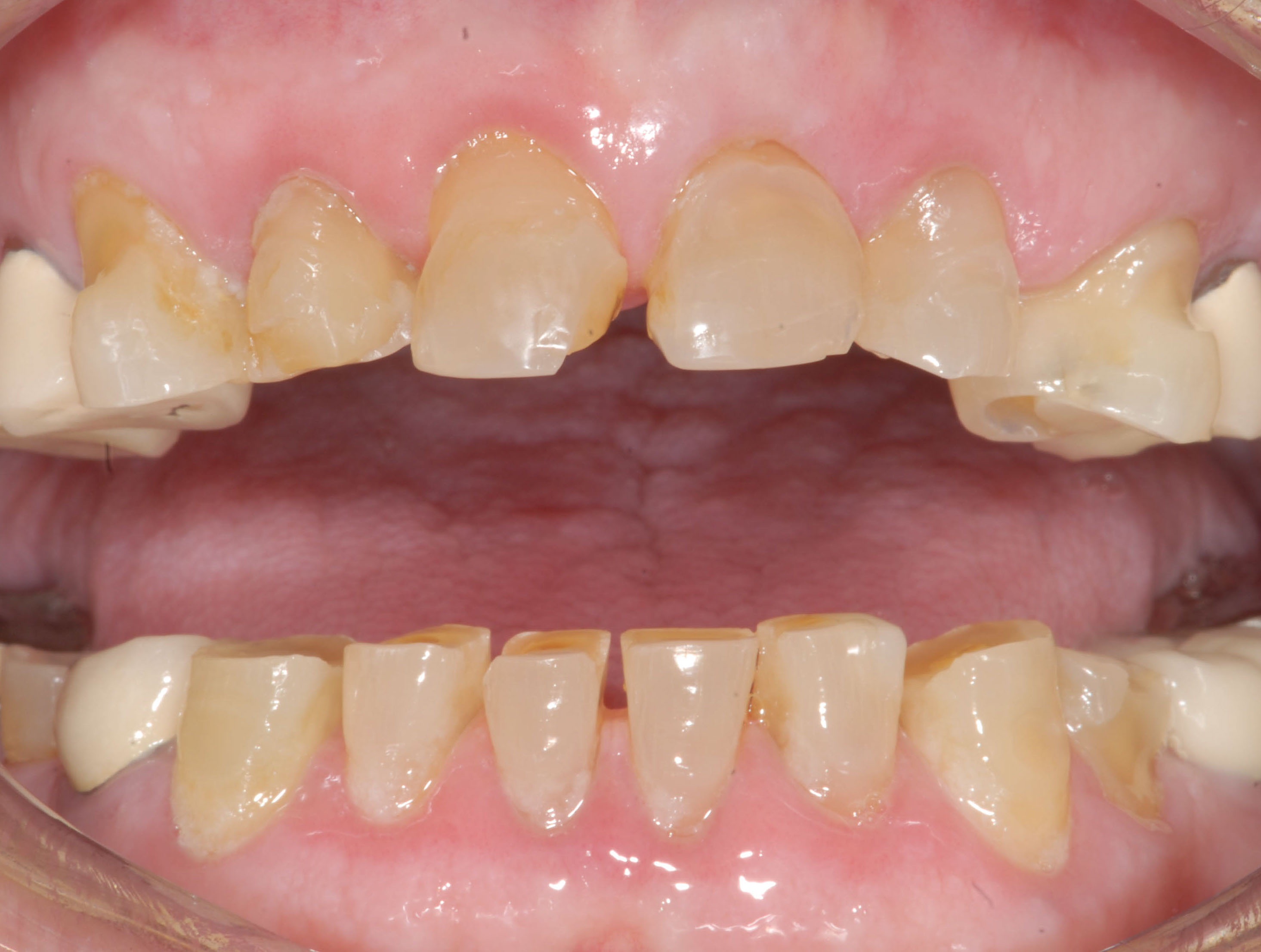
Severely worn teeth requiring full mouth restoration.
-
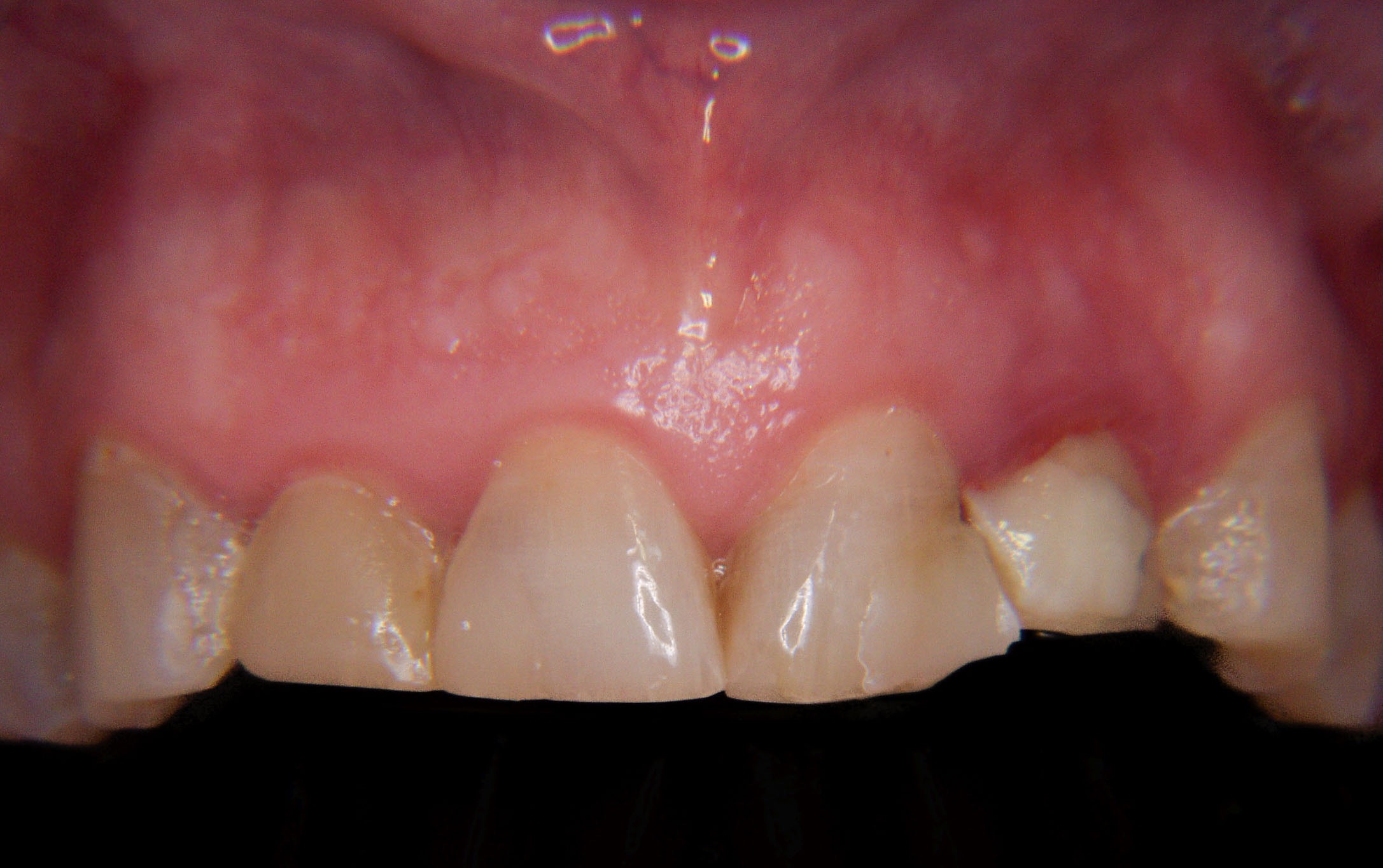
After crown lengthening to expose more tooth structure to support the planned crowns. One of the front teeth requires removal and implant replacement.
-
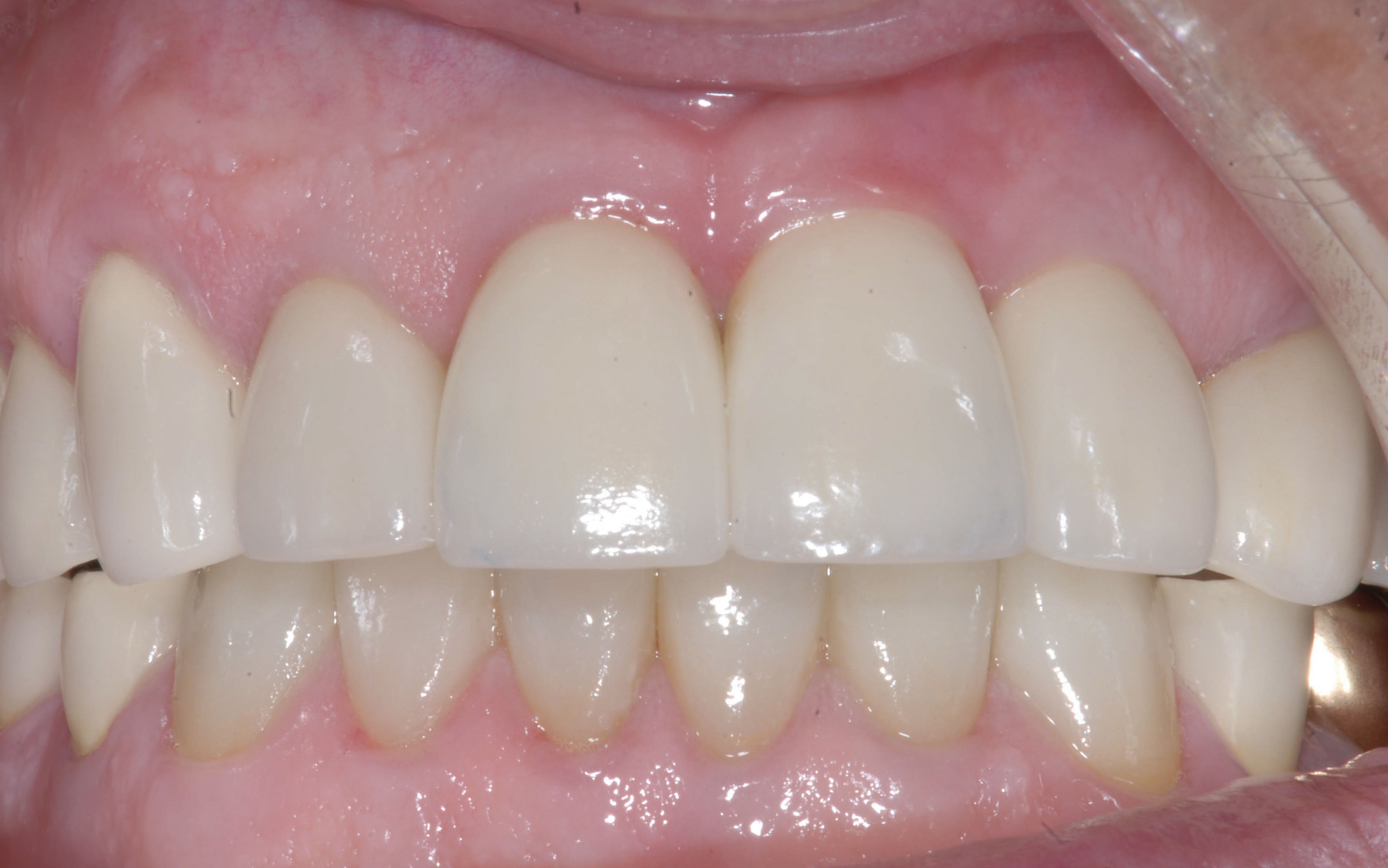
Completed reconstruction.
-
-
Severely worn teeth before treatment.
-
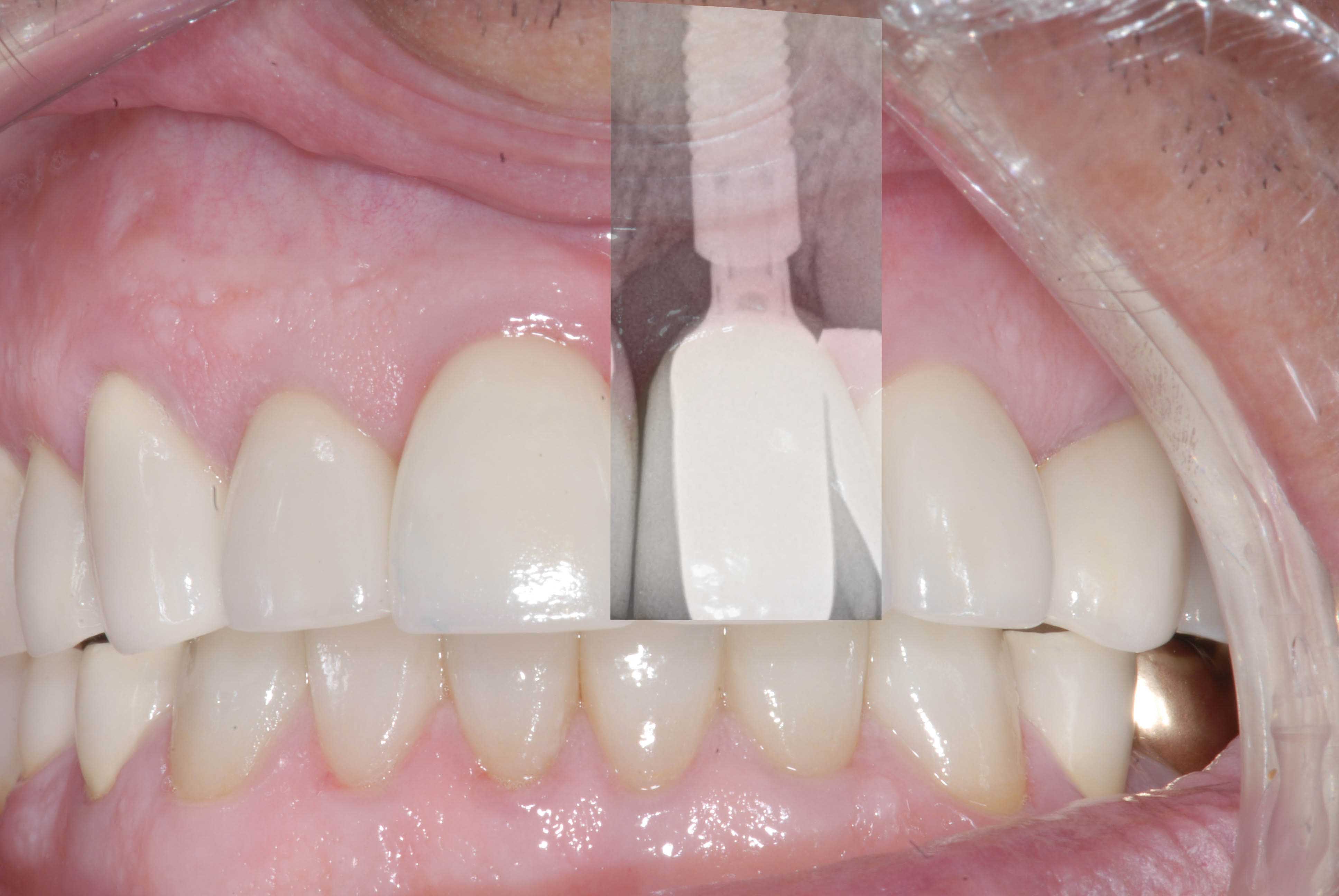
Restorations in place after crown lengthening and implant replacement of one of the front teeth.
-
Radiograph overlay of implant .
-
Congenitally missing incisor teeth. If teeth are missing the bone in that area can atrophy or diminish to an extent that an implant cannot be placed (note concavity). Bone grafts were used to increase the bone volume to allow implant placement.
-
Radiographic overlay of the implant restorations.
-
Completed case after 4-5 years in function.
-
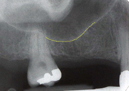
Radiograph showing sinus floor (yellow line), the sinus cavity can increase in size over time in the molar area after a tooth has been removed. Here there is insufficient vertical bone height to place an implant. A sinus elevation procedure is commonly performed to increase the bone height.
-
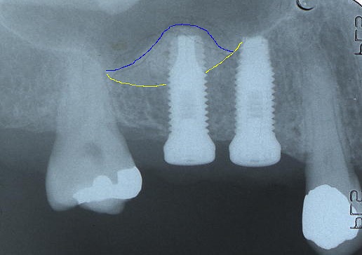
Radiograph at the time of implant placement, The sinus membrane has been elevated from the yellow to the blue line and filled with graft material to permit the implant placement. The other site has adequate bone for the implant to be placed.
-
Radiograph of the implants 2-3 years after restoration, note the new bone over the grafted implant.
-
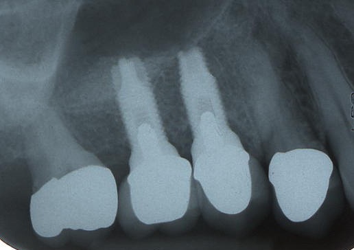
The implant restorations after 5 years, the existing molar tooth required removal due to decay.